X-Ray Imaging
Peering Inside the Body
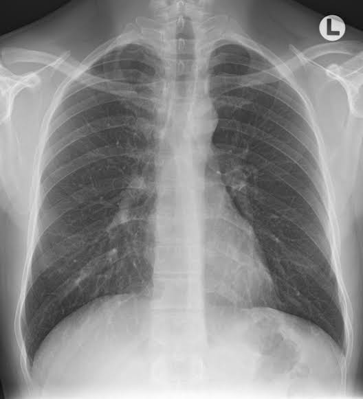
X-ray imaging, one of the most well-known and widely utilized medical imaging techniques, uses controlled amounts of radiation to visualize the internal structures of the body. This allows for the non-invasive assessment of various medical conditions.

X-rays are a form of electromagnetic radiation, similar to visible light but of higher energy. When they penetrate the body, dense structures such as bone absorb more X-rays and appear white on the X-ray image, while less dense tissues appear darker.
- Applications: X-ray imaging is essential for diagnosing and monitoring various conditions, from bone fractures to infections and tumors.
- Safety Considerations: While X-rays use radiation, the dose for most examinations is low, posing minimal risk.
- Technological Advancements: Modern X-ray machines and techniques reduce radiation exposure while ensuring high-quality images.
In the medical world, X-ray imaging remains an invaluable tool for swift and effective diagnosis, guiding treatment plans and monitoring recovery progress.
A Legacy of Medical Imaging
Since its discovery in 1895, X-ray imaging has revolutionized the field of medicine. As technology continues to evolve, X-ray systems have become more sophisticated, providing clearer images and greater insights into the human body.
